Attic Cholesteatoma Radiology

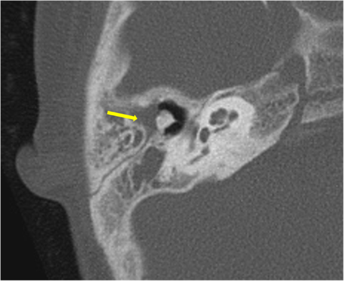
Fd Acquired Pars Flaccida Cholesteatoma Left Coronal T Bone Ct Image Shows An Atticoantral Nondependent Homogeneous Soft Radiology Image Shows Head And Neck

Cholesteatoma

Acquired Cholesteatoma Radiology Reference Article Radiopaedia Org

Mastoditis Middle Ear Head And Neck Sinusitis

Choleastoma In 2020 Middle Ear Eustachian Tube Dysfunction Ear Infection

Cholesteatoma Radiology Reference Article Radiopaedia Org
The external acoustic canal is a rare location for a cholesteatoma with an estimated incidence around 1 2 per 1 000 new otological patients.
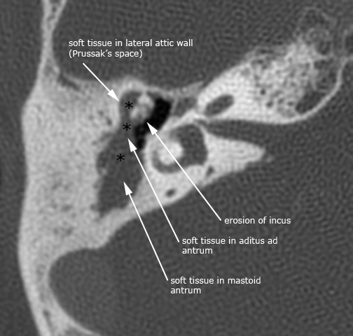
Attic cholesteatoma radiology.

Eardrums Seen In 8 Conditions Normal Eardrum Acute Otitis Media Perforation Small Perforation Attic Perforat Otitis Otitis Media Health Assessment Nursing
Hrct Imaging Of Acquired Cholesteatoma A Pictorial Review Springerlink
Pars Tensa Cholesteatoma Radiology Case Radiopaedia Org

Cholesteatoma Radiology Case Radiopaedia Org

Image Result For Scutum Erosion Facial Nerve Eustachian Tube Dysfunction Middle Ear

The Radiology Assistant Pathology

Cholesteatoma Radiology Key

Mastoditis Middle Ear Head And Neck Sinusitis
Inflammatory Ear Conditions

Ct Through The Orbits Obtained Initially Without Contrast And Then With Contrast While The Patient Performed A Valsalva Manoeuvre In The Kt Ppn
Http Pdf Posterng Netkey At Download Index Php Module Get Pdf By Id Poster Id 119105
Http Pdf Posterng Netkey At Download Index Php Module Get Pdf By Id Poster Id 115714

8 Congenital Cholesteatoma Of The Middle Ear Ento Key

A Case Of Bilateral Congenital Middle Ear Cholesteatoma

Primary And Secondary Cholesteatomas Cholesterol Granuloma And Mucocele Of The Temporal Bone Role Of Computed Tomography And Magnetic Resonance Imaging With Emphasis On Diffusion Weighted Imaging Sciencedirect

Pin On Uho

Female Reproductive System Radiology Key Radiology Female Reproductive System Reproductive System Female

5 Skull Base And Temporal Bone Table 5 1 Table 5 4 Radiology Key
Https Encrypted Tbn0 Gstatic Com Images Q Tbn 3aand9gcshxigp1cp Lksw5eyrjfubeo7r11ccyg1sj86hrxlmpnhx Awv Usqp Cau

Ct Morphological Evaluation Of Anterior Epitympanic Recess In Patients With Attic Cholesteatoma Semantic Scholar

Cholesteatoma Associated With Squamous Cell Carcinoma Of The External Auditory Canal Case Report And Literature Review Sciencedirect

Temporal Bone Radiology Key

Mbbs Doctors Atticoantral Chronic Suppurative Otitis Media Otitis Media Otitis Chronic

Mbbs Doctors Atticoantral Chronic Suppurative Otitis Media Otitis Otitis Media Chronic
Source : pinterest.com
